By Czakó Norbert, Silaghi Ciprian, Vizitiu Ramona Stela, “A smile with MIRA” Rehabilitation Centre, Cluj-Napoca, Romania, norbertczako@unzambet.com
This study is presented at The 6th IEEE International Conference on E-Health and Bioengineering – EHB 2017, Sinaia, Romania, June 22-24, 2017 and available online on IEEE Xplore.
Abstract— This case study presents the results obtained in the recovery of the brachial plexus palsy caused by transverse myelitis. The recovery of the brachial plexus palsy, whatever the etiology (traumatic, infectious or autoimmune), is a process that requires a complex and long term rehabilitation plan. A 7 year-old boy presented acute motor deficit of the left upper limb after a viral respiratory infection. The diagnosis was transverse myelitis that caused the brachial plexus palsy. We described the results obtained after 4 months of recovery with MIRA exergames and Vojta therapy.
I. INTRODUCTION
This report describes a rare case in which only the upper roots of the left brachial plexus was affected by the transverse myelitis of viral etiology and the rehabilitation program for the recovery of the motor deficit. The recovery plan consisted of MIRA exergames and Vojta therapy. The plan was based on the neurologist’s recommendations and therapeutic objectives specific to the diagnosis. The results shown here are after 4 months of therapy. The patient included in this study was diagnosed with superior brachial plexus palsy following a transverse cervical C3-C6 myelitis.
II. CASE STUDY PRESENTATION
A. Anamnesis
Seven-year-old boy is hospitalized for acute motor deficit of the left upper limb, a viral respiratory infection and anxiety.
General Examination: Good general condition, no fever, weight 38 kg, pale, excesses subcutaneous fat, good cardiorespiratory balance.
Neurological Examination: Left upper limb in adduction with internal rotation; is able to perform finger flexion/extension, hand pronation/supination; there are signs of palsy, muscular force diminished, anxiety. Spine MRI revealed myelitis, possibly viral origin.
Recommendations: Medication and physiotherapy.
B. Rehabilitation plan
After evaluation, the main physiotherapy rehabilitation objectives were established, highlighting a protocol composed of Vojta therapy, MIRA exergames and a few physical exercises given by the physiotherapist to perform at home.
Vojta therapy is a reflex locomotion method that uses hands on treatment to enable elementary patterns of movement in patients with impaired central nervous systems and loco motor system to be restored once more [6].
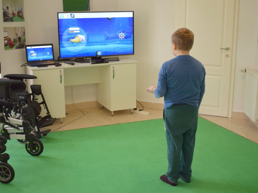
MIRA is a software platform that transforms clinical exercises in video games (exergames), enabling interaction through motion sensors (like Microsoft Kinect) and tracking exercise correctness and treatment adherence by remotely monitoring and registering movement statistics. MIRA therapy has been successfully used for the motor rehabilitation and evaluation of different neurological conditions, such as cerebral palsy or hemiparesis [7].
The exergames used in this study involved both hands, and were focused to improve range of motion, strength and coordination in order to prevent joint stiffness, to promote muscle strengthening for affected and unaffected muscles and regain a functional coordination. Initially this was done through assistive passive ROM exercises, assisted active range of motion (ROM) exercises and strengthening exercises. The program consisted in performing 7-8 exergames, 2 minutes each, 4 times per week.
First, movements were performed assisted (the non-affected arm is helping the affected arm, finger crossed at hand level), then gradually the amount of assistance was decreased (fingers on top of each other, as it can be observed in Figure 1) and when it became possible, active movements were performed, against gravity and resistance (thera-bands, weights and wrist bands were added).
C. Motor evaluation of the patient
Functional deficit evaluation has been assessed using Mallet scale for brachial plexus palsy [8] which can be used for children older than 2 years old and evaluates the active abduction and external rotation of the arm and the ability to reach out the neck, mouth and back with the hand. The result is classified in 3 stages:
• I: frozen shoulder in vicious position, holds the arm in adduction, internal rotation, extended and pronated elbow (vicious position);
• II: frozen shoulder in functional position refers to the possibility of the child to get out from the vicious position described for the value I;
• III and IV: intermediary stages;
• V: normal shoulder.
Muscular testing (strength) was measured with Medical Research Council Scale, presenting values 0 to 5, in which 0 presents no palpable contraction of the tendon, 1-2 patient can execute partial or complete movement with no gravity, 3-4 patient executes the movement against gravity, and 5 the patient maintains position against a maximal resistance.
III. RESULTS
On the first evaluation, when the patient came to our clinic he had no movement at all on his left upper limb. After 4 months of therapy we reevaluated the patient and results are presented in the table below. The biceps brachialis muscle was the first muscle that showed improvement and elbow flexion was the first movement that was possible against gravity without assistance from the non-affected arm. At this point, resistance was added by weighted balls, thera-band or weighted wrist bands.
Based on the Mallet scale and the MRCS evaluation the most of the functionality, and respectively the strength, where regained.
IV. CONCLUSIONS
Based on the results obtained so far we concluded that MIRA combined with Vojta therapy can be successfully applied in the acute stage of brachial plexus palsy. The next steps of the treatment, we plan on using MIRA only with the affected arm on all the movements used during therapy, including elbow flexion. Further down the recovery process we will apply resistance with thera-band and/or weights to all the movements performed throughout the therapy session. Initially, we combined Vojta and MIRA in our sessions but it became increasingly difficult to maintain the correct position due to the constant evasive movements. Therefore, we decided that from now on we will continue the treatment with classic therapy and MIRA only.
REFERENCES
[1] I. Albu, R.Georgia, Topographic Anatomy (Anatomie topografica), Bucuresti, 1998, ed. II, pp.66-69.
[2] Megan M. Tschudy, Kristin M. Arcara, The Harriet Lane Handbook : a Manual for Pediatric House Officers / the Harriet Lane Service, Children’s Medical and Surgical Center of the Johns Hopkins Hospital.—19th ed., pp.459-472.
[3] Michael Rubin, Acute Transverse Myelitis, MSD MAnual , New York, 2016, http://www.msdmanuals.com/professional/neurologic-disorders/ spinal-cord-disorders/acute-transverse-myelitis
[4] Varina L. Wolf, MD, Pamela J. Lupo, MD, Timothy E. Lotze, MD., Pediatric Acute Transverse Myelitis Overview and Differential Diagnosis, J Child Neurol, 2012, 27: 1426.
[5] Lindita Vata, Fadil Gradica, Detjon Vata, Edmond Pistulli, Hajrie Hundozi Hyseni, Physiotherapy Treatment of Obstetrics Brachial Plexus Palsy (OBPP) Erb – Duchenne by age group, Anglisticum Journal (AJ), Volume: 5, Issue: 9, September 2016.
[6] ***, Internationale Vojta Gesellschaft e.V., 2017 http://www.vojta.com/en/the-vojta-principle/vojta-therapy/fundamentals
[7] Moldovan, I. M., et al., Development of a New Scoring System for Bilateral Upper Limb Function and Performance in Children with Cerebral palsy Using the MIRA Interactive Video Games and the Kinect Sensor, International Conference on Disability, Virtual Reality and Associated Technologies, 2-4 September 2014, Gothenburg, Sweden.
[8] M. M. Al-Qattan and A. A. F. El-Sayed Department of Surgery and Obstetrics, King Saud University, P.O. Box 18097, Riyadh 11415, Saudi Arabia, Obstetric Brachial Plexus Palsy: The Mallet Grading System for Shoulder Function—Revisited, Hindawi Publishing Corporation BioMed Research International Volume 2014, Article ID 398121, pp.2, https://www.hindawi.com/journals/bmri/2014/398121/.
[9] Michelle A. James, MD, Use of the Medical Research Council Muscle Strength Grading System in the Upper Extremity, The Journal of Hand Surgery, Vol. 32A No. 2 February 2007, pp. 155, http://www.iprmd.org/.


